Multiplexed Imaging and Automated Signal Quantification in Formalin-Fixed Paraffin-Embedded Tissues
 Онлайн
GenomeWebinars
Sponsored by Canopy Biosciences
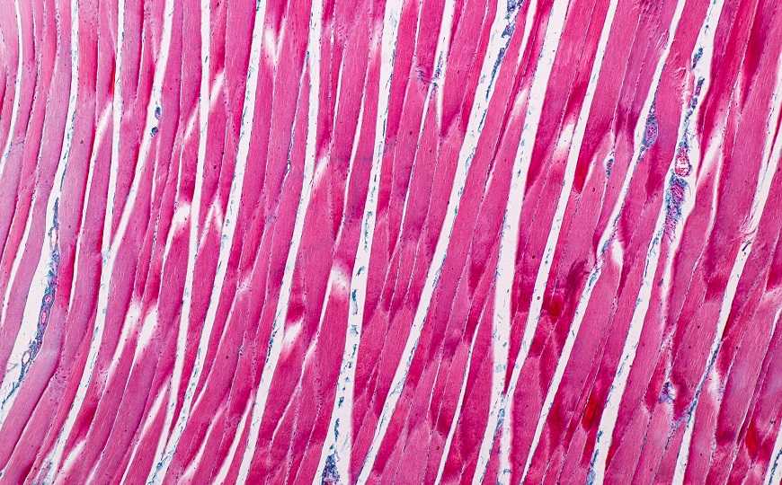
Simultaneous in situ analysis of a multitude of cells and markers is important to our understanding of tissue biology in health and disease. ChipCytometry, a user-friendly optical imaging-based technology for multiplexed staining, is a powerful tool for such analysis and can be used on both fresh-frozen and formalin-fixed paraffin-embedded (FFPE) tissue samples.
In this webinar, Sebastian Jarosch, a doctoral student at the Technical University of Munich, will describe the systematic optimization of high-plex staining and analysis of FFPE samples using ChipCytometry. Jarosch and colleagues found that the multiplexing of up to 30 markers in combination with a developed open-source workflow for signal quantification facilitated high-dimensional tissue analyses. The platform can be used for quantitative analyses of tissue composition as well as the detection of phenotypically complex rare cells, and it can be easily implemented in both routine research and clinical pathology.


 Меню
Меню


 Все темы




 0
0
